Ning Tian

E-mail:
Education:
M.D. 1982, Yichang School of Medicine; M.S. 1987, Sun Yat-sen University of Medical Science; Ph.D. 1994, SUNY at Buffalo; Postdoctoral Fellow 1994-1998, University of California, San Francisco
RESEARCH:
Synaptic plasticity
Neuronal signals are processed in vertebrate CNS through parallel synaptic pathways. These synaptic pathways are formed with distinct cellular and molecular components and, in some cases, regulated by different mechanisms during development. In many parts of CNS, including visual system, a fundamental anatomical feature of the parallel synaptic pathways is the histologically discrete laminar structure. The cellular and molecular specificity of the laminar structure appears to be a major determinant of the specific synaptic pathways. In vertebrate retina, synaptic pathways processing different aspects of visual signals are also formed with different neuronal subtypes and synaptic structures in distinct laminae. This laminar structure is not mature at birth and continues to develop during postnatal ages in most mammalian retina. The goals of our research are to understand the cellular and molecular mechanisms, which regulate the development of the retinal synaptic pathways and the formation of the laminar structure, and how these mechanisms are modulated under normal and pathological conditions. Our principal strategies are to examine retinal ganglion cell (RGC) synaptic connectivity and activity at different stages of development under normal and pathological conditions and to test specific hypotheses using appropriate transgenic animal models.
Movie 2.
Time-lapse image of a segment of RGC dendrites shows the dynamic changes of the dendritic
protrusions of RGC in developing retina.
The results of these studies provide insights to how retinal synaptic circuitry could be changed during activity-dependent synaptic plasticity. They also have important implications in how we view pathologies that affect vision during infancy and childhood.
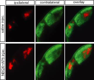 Fig 1 (dLGN).
Impaired eye-specific segregation of mouse RGC axonal projections in the dLGN due
to pharmacological blockade of spontaneous synaptic activity mediated by glutamate
receptor in retina.