Bryan W. Jones

Associate Professor of Ophthalmology & Visual Sciences
Literature & Publications
PubMed, Jones, B.W.
Cellular Neuroscience
Neurobiology of Disease
Education:
B.S. 1996, University of Utah; Ph.D. 2003, University of Utah; Post-doctoral fellow, 2003-2005 University of Utah
RESEARCH:
Retinal circuitry/connectomics and neural plasticity in retinal disease
Research: Understanding how retinal circuitry changes in disease and how it is altered from wild type conditions is critical to understanding pathogenic processes and deriving therapeutic interventions. Our work over the last few years has focused on the normal circuitry and aberrant remodeling of the neural retina and its circuitry triggered by inherited and induced retinal degenerations. This work is responsible for discovering the substantial clinical significance of negative neuronal remodeling in retinal degenerations. Ongoing work in remodeling extends to both animal disease models and human models of retinitis pigmentosa and age related macular degeneration. Future goals are to solidify our understanding of retinal circuitry as well as pathological retinal circuitry, particularly earlier in the disease process by creating complete network diagrams with rich data including classes, cell patternings, and complete connectivities. This work is fundamental for comparison and understanding of aberrant or corrupt circuitry observed in neurogenetic models as well as diseases that trigger retinal remodeling such as age related macular degeneration and retinitis pigmentosa.
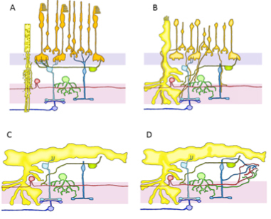
Retinal remodeling, seen in the above cartoon is a phased negative revision of neural
circuitry and topology, observed in retinal degenerative diseases. This plasticity
renders retinal circuitry corruptive of visual processing and reflects attempts by
neurons to replace lost synaptic excitation. However, this unprecedented adult plasticity
might also be exploited to rescue vision.
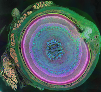 Anti-taurine, anti-glutamine and anti-glutamate assigned to red, green and blue color
channels respectively were used for this image from a 7 channel dataset. This image
shows two fundamentally important things about metabolomics: 1) Diversity of metabolomic
signatures across cell classes is extensive and in this case, it shows how complex
the retina is. 2) Within cell classes, metabolomic envelopes are very narrowly constrained
in healthy tissue.
Anti-taurine, anti-glutamine and anti-glutamate assigned to red, green and blue color
channels respectively were used for this image from a 7 channel dataset. This image
shows two fundamentally important things about metabolomics: 1) Diversity of metabolomic
signatures across cell classes is extensive and in this case, it shows how complex
the retina is. 2) Within cell classes, metabolomic envelopes are very narrowly constrained
in healthy tissue.